Feature 05 of 07
From organ to cell, in one click.
Select a structure in a 3D organ and look closer: the kidney opens its nephron, the lung its alveoli, the cochlea its organ of Corti. The scale changes; the context stays.
- Links
- 164 from organ to cell
- From
- Heart, lungs, abdomen, brain, eye and ear
- To
- Heart wall, alveoli, nephron, retina and more
- Tools
- The same labels, views and slices
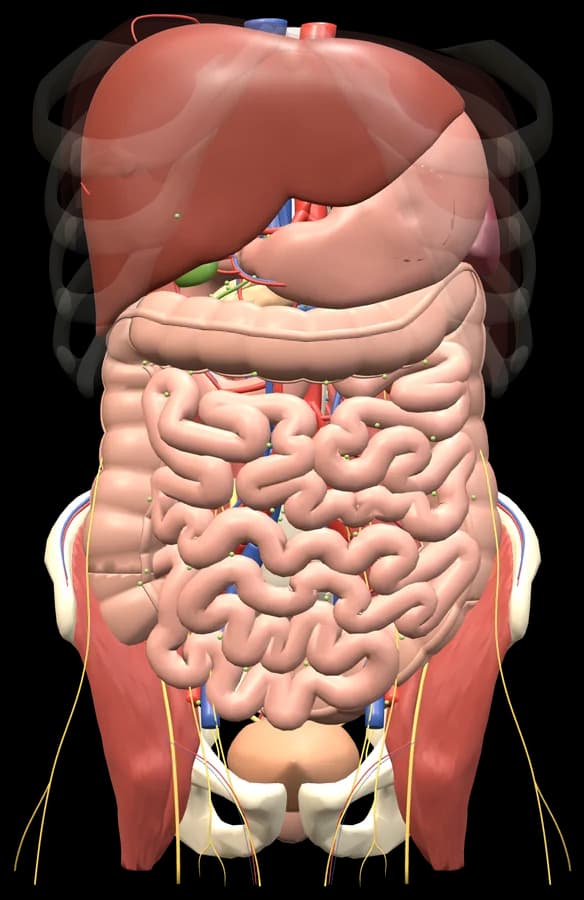
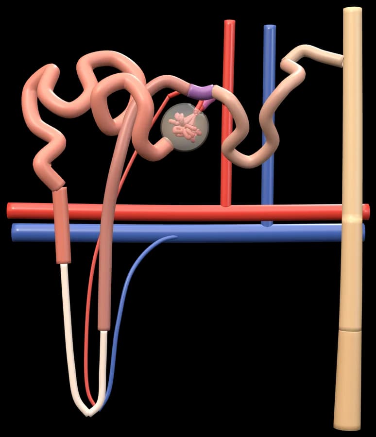
From the kidney in the abdomen to the nephron, one structure card away.
The problem
Anatomy and histology are taught apart.
- 01
Two subjects, one organ.
Gross anatomy and histology are often taught in different weeks, by different people, from different books.
- 02
Lost in scale.
A nephron is a fraction of a millimetre long. Without a link back to the kidney, it is hard to place.
- 03
Function lives in the small parts.
Filtration, gas exchange and absorption happen at the level of cells. The big picture alone cannot explain them.
How it works
Open, select, look closer.
Open an organ.
The heart, the lungs, the abdomen, the brain, the eye or the ear.
Select a structure.
Its card opens with its name and what it does.
Press Look closer.
Structures with a microscopic model show the link on their card.
Explore the close-up.
The microscopic model has its own labels, study views and, often, a real slide.
Why it helps
One atlas, every scale.
- A
164 links from organ to cell.
The heart wall and its conduction system, the alveoli, the liver lobule, the villus, the nephron, the retina and more.
- B
The same tools at every scale.
Turn, peel, slice and label the close-up just as you did the organ.
- C
Slides at the small end.
Many close-ups have a real microscope slide beside them, so you see the tissue as it really looks.
Questions
Questions, answered.
Still deciding? It is free to start, in your browser.
Which organs have a closer look?
The heart (its wall and conduction system), the lungs (the alveoli), the liver, small intestine, spleen, kidneys and adrenal glands, the brain (its pathways), the eye (the retina) and the ear (the organ of Corti), among others.
Are the microscopic models to scale?
They are enlarged so you can read them, and each model says how much in its scale note.
Is it free?
The demo models are free with Explore: seven demos: heart, arm, brain, eye, skull, glucose and DNA. Student Pro opens every model, including the microscopic ones.
Everything in Get3DMed